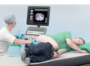
Fusion biopsy procedures combines information from diagnostic multiparametric magnetic resonance imaging (mpMRI) with real-time ultrasound imaging to target suspicious areas within the prostate for biopsy. Image-guided biopsy are being performed cognitively or supported by dedicated software. Based on the assigned ISUP grade of the collected tissue samples, the aggressiveness of the tumor, the appropriate course of action are determined.
Shortcomings of Fusion Biopsy
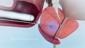
Targeting prostatic lesions with ultrasound during fusion biopsy can be challenging and requires expertise. Especially lesions located anteriorly or in the apex of the prostate are difficult to reach. Similar to systematic biopsy, Image-guided biopsy may miss small or inconspicuous tumors, leading to false-negative results or they may also sample lesser-aggressive cancerous tissue, leading to underestimation of the true aggressiveness of prostate cancer.
Fusion biopsy requires usage of local anesthesia where for in-bore biopsy only lidocaine gel is used for the procedure. Typically Image-guided biopsy is performed in combination with systematic biopsy, which accordingly leads to similar complication rates due to the large number of cores taken.
Utilizing in-bore biopsy allows for targeting the entire prostate gland and with use of near real-time MR-imaging lesions can be precisely targeted in addition to confirmation of the biopsy location.
Soteria Medical has developed a robot for in-bore MR-guided prostate interventions – based on a novel, patented motor principle.
HIGH ACCURACY
Fast and precise in-bore targeting of tumor suspicious areas within the prostate gland, based on near real-time images.
FAST PROCEDURES
Using our unique solution patients can be biopsied within a short timeframe, making the procedure cost-effective.
LESS STRESS
Due to the increased accuracy and low number of biopsy cores, the procedure is less stressful for the patient, improving the diagnostic outcome.
Please contact our experts.
