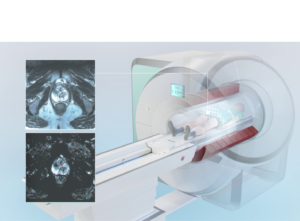
The diagnosis of prostate cancer involves several steps to determine the presence of cancer in the prostate gland and its aggressiveness. Abnormal prostate-specific antigen (PSA) blood test and digital rectal examination (DRE) are followed by either biopsy and/or a diagnostic MRI with PI-RADS scoring. Persistent suspicious cases require MR-guided targeted biopsy to establish diagnosis and the true aggressiveness of the tumor to determine the correct treatment.

Prostate cancer is the development of cancerous cells within the prostate gland. It is one of the most common cancers in men. Understanding the statistics and risk factors is vital to save lives. Early detection is crucial, as it paves the way for the right treatment.
A random systematic prostate biopsy collects 10-16 tissue samples from the prostate gland under ultrasound guidance, typically when screening abnormalities suggest possible prostate cancer.

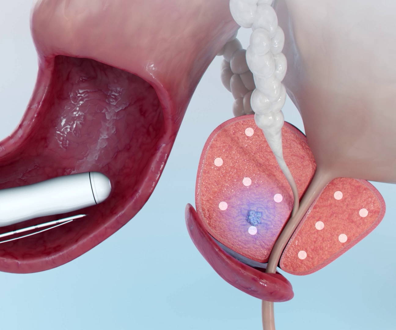
A fusion prostate biopsy, also known as MRI-ultrasound fusion biopsy, is a diagnostic procedure used to target a cancer suspicious lesion within the prostate gland by fusing MRI diagnostic images with ultrasound guidance.

In-bore MR-guided biopsy is the most advanced diagnostic procedure using real-time MR imaging to target tumor suspicious lesions within the prostate gland and obtain tissue samples. By using the most sensitive imaging method combined with remote controlled navigation this allows for the most accurate diagnosis of prostate cancer.
Selecting the appropriate biopsy method is crucial, and it depends on the patient’s medical history and/or diagnostic imaging. There are various biopsy methods, each with its own advantages and disadvantages. The choice of method plays a significant role in accurate diagnosis.